
-
What is Brain-Computer Interface (BCI) ?
Rehabilitation medicine is one of the critical issues for cerebropathia, paraplegia or brain nerve damaged patient in terms of being able to control motor ability or outer circumstance. A brain-computer interface (BCI), often called a mind-machine interface (MMI), or sometimes called a direct neural interface or a brain-machine interface (BMI), is highlighted as a direct communication pathway between the brain and an external device. BCIs are often directed at assisting, augmenting, or repairing human cognitive or sensory-motor functions.
-
Our Aim
Our team aim to decode the intention using brain signal. We are trying to understand brain activity and associated electrocorticogram (ECoG) signal, and to figure out epidural ECoG feasibility for minimal invasive recording technique. In past few years, we tried to decode the non-human primate (NHP) movement intention such as saccadic movement or bimanual movement, using epidural ECoG. Lately, we aim to predict the trajectory of NHP bimanual movement for BCI using machine learning algorithm including deep learning.
-
Topics
- Bimanual arm movement tracking in non-human primates
- Investigation of the functional neural connectivity
- Deep learning based BCI
- Screw ECoG development
- Development of Temporal Interference stimulation technique using high frequency (>1kHz) current

-
Recent Papers
Improved prediction of bimanual movements by a two-staged (effector-then-trajectory) decoder with epidural ECoG in nonhuman primates
(Hoseok Choi et. al., Journal of Neural Engineering, 2017)
It is well known that the bimanual brain state is different from the unimanual one. Conventional methodology used in unimanual studies does not take the brain stage into consideration, and therefore appears to be insufficient for decoding bimanual movements. So the monkeys performed three types of arm movement tasks: left unimanual, right unimanual, bimanual. To decode these movements, we used a two-staged decoder, which combines the effector classifier for four states (left unimanual, right unimanual, bimanual movements, and stationary state) and movement predictor using regression. The results showed that decoding performance for bimanual movements were improved compared to the conventional method, which does not consider the effector, and the decoding performance was significant and the stable over a period of four months

A fronto-parietal network, comprised of the posterior parietal cortex (PPC) and the dorsal premotor cortex (PMd) has been proposed to be involved in planning and guiding movement. However, the issue of how the network is expressed across the bilateral cortical area according to the effector's side remains unclear. In this study, we tested these questions using electrocorticographic (ECoG) recordings in non-human primates using a simple visual guided reaching task that induced a left or right hand response based on relevant cues provided for the task. The findings indicate that that right hemisphere dominant network patterns in which the right PMd was strongly coordinated with bilateral PPC immediately after presentation of the movement cue occurred, while the coherence with the left PMd was not enhanced. No difference was found in the coherence pattern between the effector’s side (left hand or right hand), but the strength of coherence was different, in that animals showed a higher coherence in the right hand response compared to the left. These support the conclusion that right hemisphere dominance in long-range phase synchrony in the 10–20 Hz low beta band is involved in motor preparation stage, irrespective of the upcoming effector’s side.
Right hemisphere dominance in neural connectivity within fronto-parietal networks in non-human primates during a visual reaching task
(Jeyeon Lee et. al., in preparation)

Sex differences of cognitive load effects on object-location binding memory
(Jinsick Park et. al., Biomedical Engineering Letters, 2017)

In this study, we investigated where the sex differences of object-location binding memory performance were influenced by the cognitive load. We used the fractal objects version of the ‘What was where?’ task to measure object memory, location memory and object-location binding memory. Cognitive load was controlled by task difficulty presented two sessions: one session randomly displayed three or four fractal objects (Session 34) and the other session four or five objects (Session 45). The results showed that females outperformed males on object-location binding memory. Interestingly, even when the four object trials were compared between Session 34 and Session 45, in which we believed that the level of difficulty was similar while cognitive load varied, the swap error of males was significantly increased in Session 45 compared to females. In conclusion, there may be sex differences in object-location binding memory and the males could be more sensitive about the cognitive load than females.
Neural correlates of spatial and nonspatial attention determined using intracranial electroencephalographic signals in humans
(Taekyung Kim et. al., Human Brain Mapping, 2016)
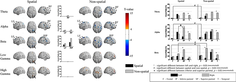
We investigated the neural correlates of spatial and nonspatial attention in humans using intracranial electroencephalography. The topography and number of electrodes showing significant event-related desynchronization (ERD) or event-related synchronization (ERS) in different frequency bands were studied in 13 epileptic patients. Performance was not significantly different between the two conditions. Spatial attention involved right-lateralized activity that was maximal in the right superior parietal lobule (SPL), whereas nonspatial attention involved wider brain networks including the bilateral parietal, frontal, and temporal regions, but still had maximal activity in the right parietal lobe. Within the parietal lobe, spatial attention involved ERD or ERS in the right SPL, whereas nonspatial attention involved ERD or ERS in the right inferior parietal lobule. These findings reveal that common as well as different brain networks are engaged in spatial and nonspatial attention.